Home

ct scan of vocal cords

CT scan: heterogeneously enhancing mass lesion on the false vocal cords. |  Download Scientific Diagram
CT scan: heterogeneously enhancing mass lesion on the false vocal cords. | Download Scientific Diagram
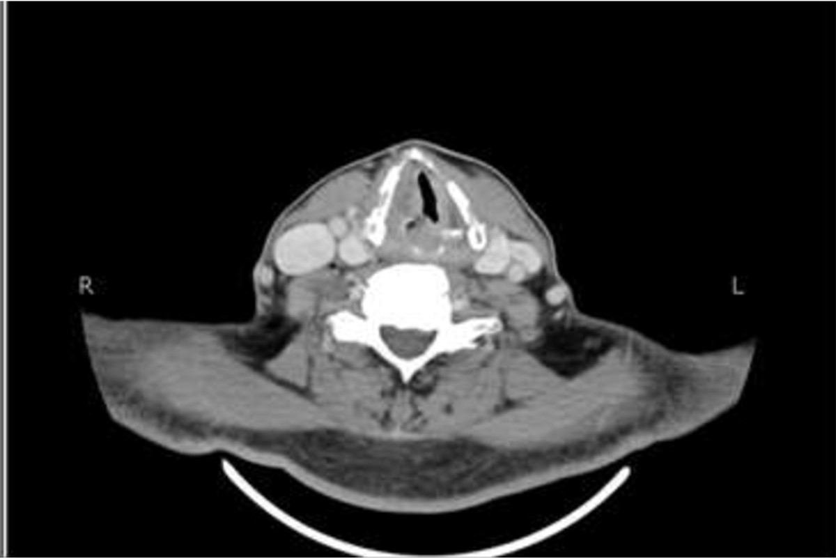
Vocal cord paralysis: anatomy, imaging and pathology | Insights into Imaging  | Full Text
Vocal cord paralysis: anatomy, imaging and pathology | Insights into Imaging | Full Text

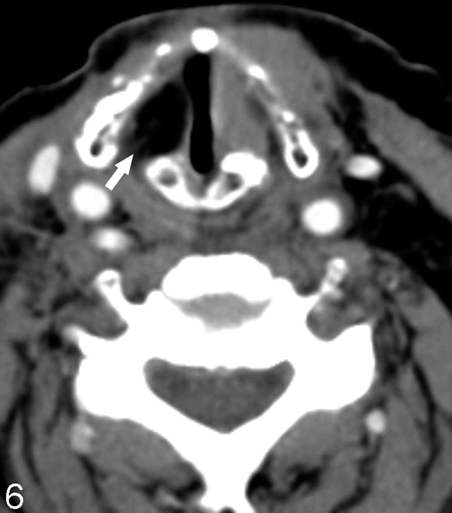
CT Assessment of Vocal Cord Medialization | American Journal of  Neuroradiology
CT Assessment of Vocal Cord Medialization | American Journal of Neuroradiology

Vincristine-induced vocal cord palsy and successful re-treatment in a  patient with diffuse large B cell Lymphoma: a case report | BMC Research  Notes | Full Text
Vincristine-induced vocal cord palsy and successful re-treatment in a patient with diffuse large B cell Lymphoma: a case report | BMC Research Notes | Full Text

EPOS™
EPOS™

Imaging of larynx - YouTube

CT Assessment of Vocal Cord Medialization | American Journal of  Neuroradiology
CT Assessment of Vocal Cord Medialization | American Journal of Neuroradiology

Normal anatomy of the false and true vocal cords on computed tomography

Axial CT scan obtained at the level of the vocal cords during quiet... |  Download Scientific Diagram
Axial CT scan obtained at the level of the vocal cords during quiet... | Download Scientific Diagram

Paralyzed Vocal Cord, CT - Stock Image - C007/5844 - Science Photo Library

Left vocal cord lesion - squamous cell carcinoma | Radiology Case |  Radiopaedia.org
Left vocal cord lesion - squamous cell carcinoma | Radiology Case | Radiopaedia.org

Normal appearance of the vocal cords. a Axial CT images during quiet... |  Download Scientific Diagram
Normal appearance of the vocal cords. a Axial CT images during quiet... | Download Scientific Diagram

Axial (a) and sagittal (b) CT of the neck with contrast demonstrate a... |  Download Scientific Diagram
Axial (a) and sagittal (b) CT of the neck with contrast demonstrate a... | Download Scientific Diagram

Using CT to Localize Side and Level of Vocal Cord Paralysis | AJR

CT Scan of the Larynx: Overview, Laryngeal Embryology and Anatomy,  Laryngeal Neoplasms
CT Scan of the Larynx: Overview, Laryngeal Embryology and Anatomy, Laryngeal Neoplasms

Unilateral Vocal Cord Paralysis: A Review of CT Findings, Mediastinal  Causes, and the Course of the Recurrent Laryngeal Nerves | RadioGraphics
Unilateral Vocal Cord Paralysis: A Review of CT Findings, Mediastinal Causes, and the Course of the Recurrent Laryngeal Nerves | RadioGraphics

Just say 'eee' for neck CT | AuntMinnie

CT Scan of the Larynx: Overview, Laryngeal Embryology and Anatomy,  Laryngeal Neoplasms
CT Scan of the Larynx: Overview, Laryngeal Embryology and Anatomy, Laryngeal Neoplasms

Larynx CT Imaging Guide Complete Guide - Medical Professionals

ijopl-6-57-g007.jpg
ijopl-6-57-g007.jpg

Using CT to Localize Side and Level of Vocal Cord Paralysis | AJR

a) Axial CT image shows the normal appearance of the true vocal folds:... |  Download Scientific Diagram
a) Axial CT image shows the normal appearance of the true vocal folds:... | Download Scientific Diagram

Normal larynx. (a) Axial CT scan shows the normal appearance of the... |  Download Scientific Diagram
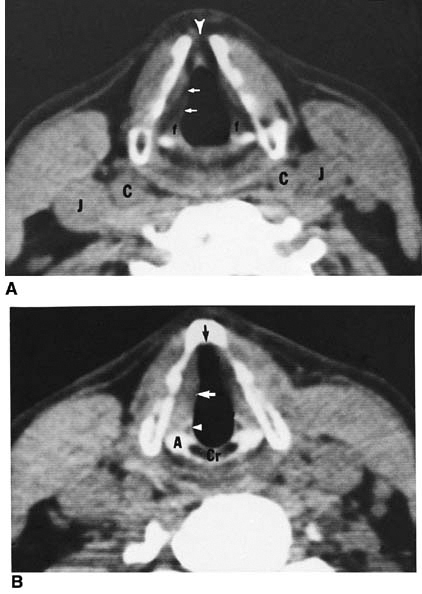
Normal larynx. (a) Axial CT scan shows the normal appearance of the... | Download Scientific Diagram